-
Research
3D bioprinting: transforming medical images into human tissue
Share this:


Mayo Clinic researchers are using technology to produce tissue models of different body parts to study damaged or diseased tissues and organs. They envision a day when a 3D bioprinter could mold living cells into a therapy or cure for complex disorders.
"3D bioprinters are just like the 3D printers that print small plastic toys or parts. Instead of using hard plastics or metals to construct a part or prototype, 3D bioprinters use biocompatible materials containing living cells to print three dimensional structures of tissue that could be used to improve human health," says Kevin Dicker, Ph.D., a bioprinting expert and a process development staff scientist for the Center for Regenerative Biotherapeutics in Arizona. "Bioprinters are tools to accelerate research in the field of tissue engineering."
3D bioprinting holds intrigue among researchers for its potential to study disease progression and screen new treatments for conditions such as end-stage organ failure, cartilage defects and atopic dermatitis, a skin disease also known as eczema. Dr. Dicker and the process development team are working to establish standard operating procedures for biomanufacturing tissue for testing in early-stage clinical trials. This pioneering work at Mayo Clinic is aimed at integrating tissue engineering into clinically relevant therapies that could be studied in clinical trials.
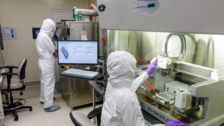
Dr. Dicker utilizes a 3D bioprinter to study tissue engineering.
Mayo Clinic's Center for Regenerative Biotherapeutics supports research to advance tissue engineering and cell and gene therapies to clinical care for patients with rare and complex diseases.
Bioprinting with living cells
The 3D bioprinter utilizes a digital blueprint of a design from medical imaging, such as MRI or CT scans, and then gets to work. This powerful, high-tech tool precisely places bioinks composed of living cells, hydrogels, biomaterials and growth factors in a layer-by-layer fashion. The final 3D tissue model it produces can mimic the structure, mechanics and physiology of human organs, muscle or cartilage.
"You can formulate a bioink composed of gelatin-like materials which can be printed into a desired shape. In addition, we can encapsulate living cells into that gelatin-like bioink," says Dr. Dicker. "This type of tissue engineering is an emerging technology in regenerative medicine that has the potential to transform laboratory research and clinical practice by bioengineering replacements for damaged or diseased tissue."
Bioprinting organs
The complex tissue structures that come from the 3D bioprinter have allowed researchers to study ways to bioprint human organs. Mayo Clinic has developed the capability of bioprinting skin to mimic inflammatory skin disease. This bioprinted skin model is being studied in the lab of Saranya Wyles, M.D., Ph.D., to test treatments and understand disease progression for conditions such as atopic dermatitis (eczema).
"We have created the first-in-kind human skin model with functional epidermis and dermis, recapitulating human skin. This 3D model gives us a humanlike model for studying inflammatory skin conditions. It accurately recreates patient-specific disease and provides the ability to test new therapies," says Dr. Wyles.
Besides its use for disease models, this emerging technology is being explored for manufacturing human tissue and organs.
"The ultimate goal is to someday be able to print organs and tissue on demand. However, we aren’t quite there yet," says Dr. Dicker. "We hope to advance this technology as a solution to the global shortage of donor organs. If we are able to bioprint functional kidneys, for example, that would be a huge relief on the healthcare system."
Bioprinting implants
In Arizona, the research team of David Lott, M.D., is developing 3D bioprinted implants for the larynx or trachea. The implants could be used to replace damaged or diseased portions of the organ while maintaining the healthy tissue.
"The 3D bioprinter has the ability to construct complex tissue which has both stiff cartilage and soft tissue regions. This enables us to print vocal folds, soft tissue responsible for vocalization, and the surrounding cartilage within the larynx," says Dr. Dicker. "We hope to develop a 3D bioprinted implant for patients who need a part of their larynx removed because of disease or trauma."
Challenges to bioprinting
While 3D Bioprinting holds a high potential, Mayo Clinic and other research institutions still have challenges to overcome. In order for a bioprinted organ to function, it must have a connection to blood, oxygen and nutrients. Researchers have struggled to grow a network of capillaries and blood vessels in the bioprinted structures at scale that would deliver those vital elements. Another challenge is how to integrate the bioprinted tissues with the human body while preventing rejection of the implant.
3D bioprinting has the potential to transform healthcare, but more years of research are needed to fully understand and apply this technology.
###
Related stories:
Electrospinning biotherapies of tomorrow
Bringing regenerative technology of the future to patients today


