-
Featured News
One-of-a-Kind Compact 3T MRI Scanner Installed for Research
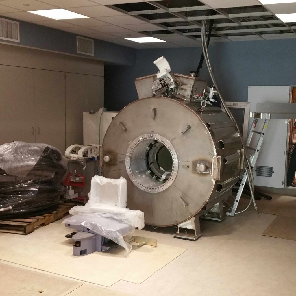
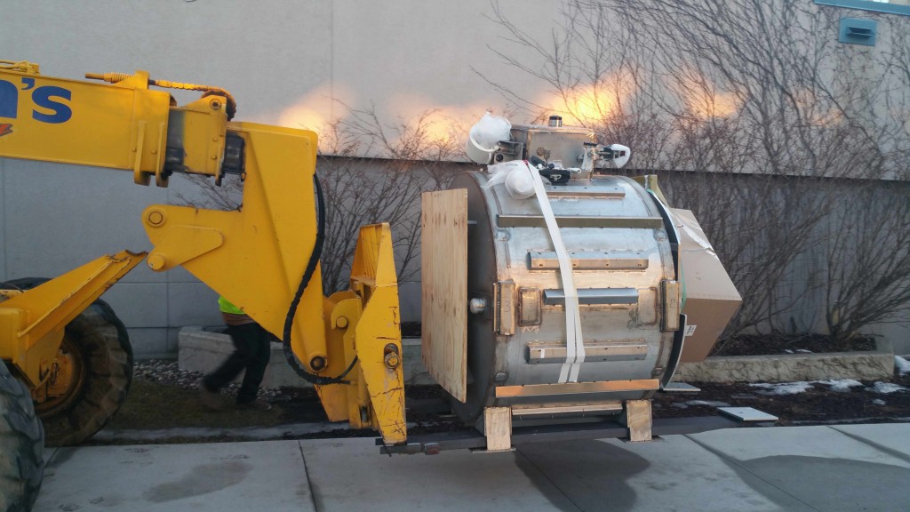
Mayo Clinic on Saturday, Feb. 20, installed a new one-of-a-kind compact 3-Tesla MRI scanner in the Charlton North Building on Mayo's Rochester campus.
The installation has been eight and a half years in the making, with teams from both Mayo and the GE Global Research Center working under a National Institutes of Health Bioengineering Research Partnership grant to develop the machine that will be housed at Mayo Clinic. A great deal of teamwork went into the development, construction and fine-tuning of the machine, and even more will go into determining the full scope of what physicians are able to do with the machine to improve patient outcomes and experiences, says Matt Bernstein, Ph.D., a medical physicist at Mayo Clinic.
MEDIA CONTACT: Ethan Grove, Mayo Clinic Public Affairs, 507-284-5005, newsbureau@mayo.edu.
The scanner will be used for conducting research with patient volunteers, as physicians and physicists look for ways to improve diagnoses and treatments of focal diseases, such as gliomas, meningiomas, stroke, aneurysm and congenital anomalies; and diffuse diseases, such as Alzheimer’s, multiple sclerosis, psychiatric disorders, leukoaraiosis and hydrocephalus.
Vital to the research program’s success are the patients who are volunteering to participate, says John Huston III, M.D., a neuroradiologist at Mayo Clinic.
“We are incredibly lucky for the generosity of our patients, that they are willing to have additional testing to help others in the future,” says Dr. Huston. “Many times, if you have a long-term problem, say multiple sclerosis, it could be that the work that we do ends up helping, and they benefit. But in general, a lot of what we’re going to do with this group of patients, it will not help them directly. Instead, it is helping to move science forward, rather than improve or impact their individual care.”
Innovative methods developed at Mayo for thecompact 3T scanner will have direct benefits for Mayo's Proton Beam Therapy Program. The technical challenge of improving geometrical accuracy for the compact 3T MRI and MRI-based treatment planning for proton beam therapy are closely related, and Dr. Bernstein says his team has been working to improve the correction that reduces distortion near the edge of scanner images.
“Some of the technology we’ve developed here spins off directly to benefit the radiation therapy planning at the Proton Beam, so we’re very excited about that,” Dr. Bernstein says.
The opportunities for multidisciplinary collaboration are part of the appeal, Dr. Bernstein says. Already, physicians who are focused on pediatrics, musculoskeletal, radiation therapy, deep-brain stimulation and Alzheimer’s disease are hopeful that the scanner’s unique capabilities will bring new insights and breakthroughs to their fields.
After the completion of the research involving 300 initial head scans, the team will explore other opportunities for the machine.
“From the beginning, we also thought the compact 3T would be useful for imaging extremities, including feet, knees and wrists,” Dr. Bernstein says. “That does increase the reach of the scanner quite a bit. Compact MRIs could handle nearly half of the clinical MRI exams we see at Mayo.”
Dr. Huston adds: “Now when we move into the next phase, there is a greater likelihood that we will be seeing things that can help individual patients. For instance, with elastography for focal diseases, we’re able to tell the surgeon before surgery if the tumor is soft or hard, and adherent to the surrounding brain or not, and that’s something that’s never been able to be done before.”
The final installation and calibration of the new scanner will be completed over the next month, with patient studies expected to begin early this spring.


