-
Research
3D-bioprinted human skin model expands options for preclinical research
Share this:





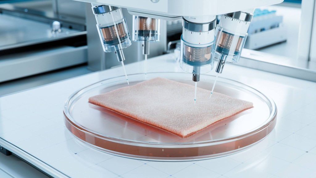
In a laboratory at Mayo Clinic, a machine that looks strikingly similar to a desktop printer is quietly reshaping the future of dermatology. Instead of ink, it dispenses living human cells. Instead of paper, it builds tissue — layer by layer — replicating one of the body's most complex organs: skin.
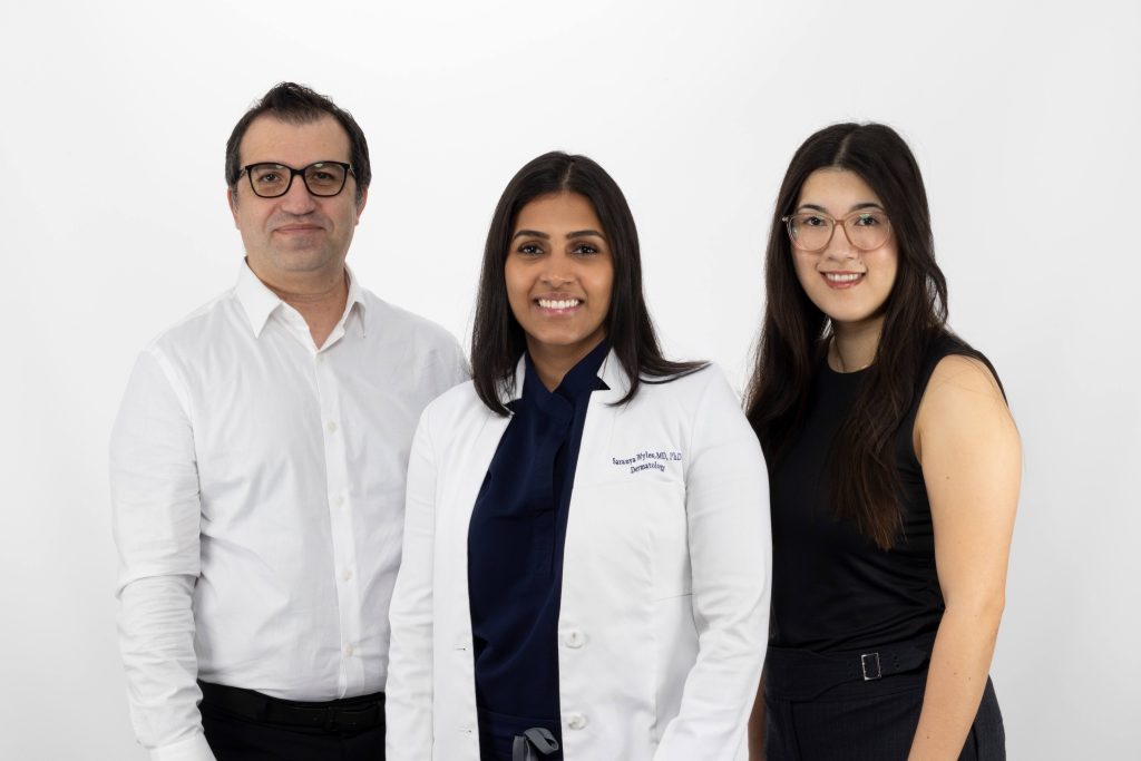
For Saranya Wyles, M.D., Ph.D., a dermatologist and researcher at Mayo Clinic, the journey into 3D bioprinting began not with an ambitious plan to reinvent tissue engineering, but with a practical problem. Her team needed a better way to test new therapies.

"We were trying to find a preclinical model to develop an FDA application," Dr. Wyles explains. Traditional approaches rely heavily on animal testing, but skin biology varies widely across species. Even commonly used preclinical models fall short when it comes to mimicking human skin conditions such as eczema.
At the same time, alternatives such as donated human skin samples — often surgical waste — can only survive for a few days in the lab. That limitation makes it difficult to study chronic diseases or long-term treatment effects.
Faced with these constraints, Dr. Wyles and her team asked a bold question: What if they could build human skin from scratch?
From printer to patient-specific model
The answer took shape through 3D bioprinting, an emerging technology that uses "bioinks" — mixtures of living cells and supportive materials — to construct tissues.
The concept sounds deceptively simple. Much like a standard printer uses different color cartridges, a bioprinter uses different cell types. In the case of skin, that includes fibroblasts, keratinocytes and melanocytes — the essential building blocks of the dermis and epidermis layers of the skin.
But translating that concept into living tissue proved anything but simple. Early attempts produced structures that looked nothing like real skin. "It was like a cartoon version," Dr. Wyles recalls. "Not even close."
What followed was nearly a year of intensive troubleshooting, bringing together clinicians, biomedical engineers and tissue engineers in a collaborative effort. The challenge wasn't just printing cells — it was keeping them alive, functional and organized.
Each cell type has its own needs, from nutrients to mechanical conditions. "It's like roommates," Dr. Wyles says. "They all want different things."
The team had to design new culture systems, optimize materials and even fine-tune the physical "stretch" of the printed tissue to mimic natural skin. These details matter. Skin's elasticity affects everything from wound healing to itch and aging. Gradually, iteration by iteration, the model improved.
Building skin, layer by layer
Today, the process resembles a carefully choreographed construction project. First, the printer lays down the dermis — the deeper layer of skin — using fibroblasts embedded in a collagen scaffold. After several days of maturation, the epidermis is printed on top, forming the outer protective layer.
The result is a structured tissue that mirrors key features of human skin, including stratified layers and pigment-producing cells. Crucially, the model uses entirely human-derived components, including a plant-based recombinant collagen that avoids the variability and immune risks associated with animal-derived materials.
The printed tissue can survive for weeks — far longer than traditional skin explants — allowing researchers to study disease progression and treatment responses over time. And because it's printed, it can be replicated with remarkable consistency.
"No two preclinical models are exactly the same," Dr. Wyles notes. "But we can print hundreds of nearly identical samples."
Improving how therapies are tested
The implications of this technology extend far beyond the lab bench. Preclinical testing has long been a cornerstone of drug development, yet it remains an imperfect predictor of how therapies will perform in humans — nearly 90% of drugs that succeed in preclinical testing ultimately fail in clinical trials.
Bioprinted human skin models offer a promising alternative — one that is not only more biologically relevant, but also faster, more scalable and more ethical.
"From both an ethical and a financial standpoint, the difference is enormous," Dr. Wyles says.
Regulators are beginning to take notice. The Food and Drug Administration has already engaged with the Mayo Clinic team on the technology, reviewing data demonstrating that the printed skin can replicate both the structure and function of human tissue.
That includes side-by-side comparisons of patient skin and lab-grown models, showing similar cellular architecture and biological responses. As a result, the model has been recognized as a promising alternative in certain preclinical contexts — potentially reducing the need for large animal studies.
Toward personalized and precision dermatology
Beyond its applications in preclinical research, the technology opens the door to a more personalized approach to medicine. Researchers are now developing ways to create patient-specific "maps" of skin by analyzing biopsies at a molecular level. These maps capture everything from cell distribution to markers of aging, such as cellular senescence.
Using this information, the team can program the bioprinter to recreate an individual's skin in the lab.
"We can essentially print your skin based on your own biological blueprint," Dr. Wyles says.
That capability could transform how treatments are developed and tested — allowing scientists to evaluate therapies on models that reflect specific patients, populations or conditions. It also has implications for studying aging, pigmentation and diseases such as atopic dermatitis, which can vary widely across individuals and skin types.
Expanding complexity
The current models represent a significant advance, but they are only the beginning. Human skin is a highly complex organ, responsible not just for protection, but also for sensation, temperature regulation and immune function. To more fully replicate that complexity, the Mayo team is working to add new features to their printed tissues.
Future versions may include blood vessels, immune cells and nerve structures — elements that would enable even more realistic modeling of disease and drug response. Researchers also are developing pigmented models to better represent diverse skin tones, addressing a long-standing gap in dermatologic research.
"Skin of color is an area where we need better models," Dr. Wyles says.
From innovation to impact
As interest in the technology grows, so does demand. Pharmaceutical and cosmetic companies are already exploring collaborations, drawn by the potential to accelerate product development while reducing reliance on animal testing.
To meet that demand, Mayo Clinic is working to scale the technology through collaborations and licensing, while continuing to refine the science.
At the same time, Dr. Wyles is mindful of the broader mission.
"We want to democratize this," she says. "The goal is to make it accessible so more people can use it, test more therapies and ultimately get treatments to patients faster."
A new frontier in regenerative medicine
The potential applications extend well beyond drug testing. Bioprinted skin could one day be used in regenerative medicine — for example, creating grafts for burn victims or patients with chronic wounds. It may also serve as a platform for studying aging and developing interventions to improve skin health over time.
For now, the focus remains on refining the models and expanding their capabilities.
"We're really just at the beginning of what this platform can do," Dr. Wyles says.