-
Research
Advancing ultrasound microvessel imaging and AI to improve cancer detection
Share this:


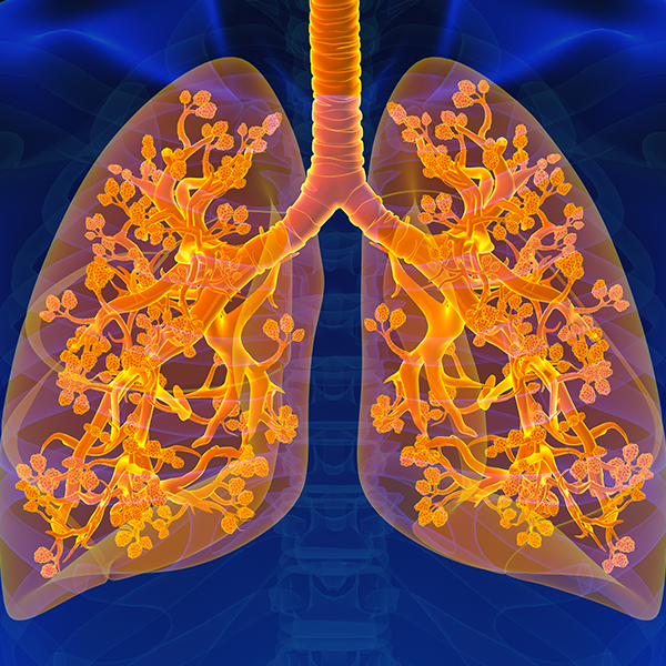
Ultrasound — a technology that uses sound waves to produce an image — is commonly used to monitor the development of a baby as it grows inside its mother. But ultrasound imaging also can be used to investigate suspicious masses of tissue and nodules that may be cancerous.
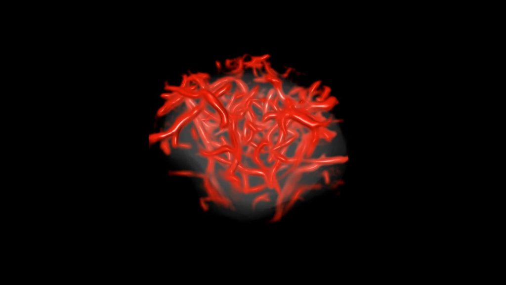
Tumors consist not only of cancer cells but also a matrix of small blood vessels, or microvessels, that cannot be seen in the images produced by conventional ultrasound machines. To solve this problem, physician-scientist Azra Alizad, M.D., and biomedical engineering scientist Mostafa Fatemi, Ph.D., teamed up at Mayo Clinic to design and study a tool that may improve the resolution of ultrasound imaging. As demonstrated in research findings, they developed high-resolution ultrasound imaging software, compatible with many ultrasound machines, that could exponentially improve both the detail and quality of images.
The investigational software, which they've named quantitative high-definition microvessel imaging (q-HDMI), has been evaluated to capture high-resolution 2D and 3D images of microvessels as small as 150 microns, roughly twice the width of a human hair.
"If we can visualize and capture the microvessel in the earliest stages of cancer, we can better diagnose and treat it earlier, which improves the outcome for the patient"
- Azra Alizad, M.D.

Artificial intelligence helps detect what we cannot see
In addition, the researchers identified a series of biomarkers representing specific characteristics of tiny
vessels, such as shape, pattern, irregularity and complexity, and packaged them into an algorithm that can sort the image data into benign or malignant masses.

"This technology provides a quantitative value that shows the probability of malignancy ... It's a tool to extract information in a way that can be useful to clinicians."
- Mostafa Fatemi, Ph.D.
Applying it in practice
In a clinical study, the researchers showed that their new q-HDMI tool combined with artificial intelligence (AI) was able to detect a very small, 3-millimeter-wide, malignant breast cancer mass comprised of miniscule vessels in a 40-year-old woman. Finding and treating cancer lesions when they are this size before they spread, or metastasize, can be lifesaving.
"The questions in radiology really are: benign or malignant? And if it's suspicious, how concerned should we be?" Dr. Fatemi says.
To help radiologists answer these questions, the researchers applied their software and algorithm to further analyze suspicious breast masses from 521 patients who already had received conventional ultrasound imaging. The results were astounding. The new technology yielded a nearly 100% rate of accuracy in determining malignant versus benign masses, regardless of the tumor size.
More recently, the researchers analyzed thyroid nodules from 92 patients. Thyroid nodules are common and it is often hard to differentiate between cancerous and non-cancerous ones through imaging. Yet the prevalence of thyroid cancer has increased over the last few decades.
"With conventional ultrasound images, physicians can diagnose whether a thyroid nodule is benign or malignant with only about 35-75% accuracy," Dr. Alizad says. That's why physicians often opt to perform thyroid biopsies, which enable them to more definitively determine if a thyroid nodule is a cause for concern.
The researchers identified 12 biomarkers that can differentiate benign from malignant thyroid tissues. They programmed the AI-powered algorithm with these biomarkers, which sorted the images and had an 84% rate of accuracy. These results were published in the journal Cancers and highlighted by the National Institutes of Health.
"If it is cancer, we definitely want to know that. But if we can determine if a thyroid nodule is benign without even having to do a biopsy, that is even better as it spares the patient from the financial and physical burdens associated with an unnecessary benign biopsy," Dr. Alizad says.
The researchers see that their quantitative tool could be particularly useful in parts of the world where there is limited expertise and resources, such as rural areas and developing countries.
They also are collaborating with oncologists to enable them to use the q-HDMI tool to better monitor the effectiveness of cancer treatments and help them adjust therapies for individual patients in real time.