-
Cancer
Exploring advanced breast cancer screening options
Share this:


Mammograms have been a standard screening for breast cancer since the 1970s. They have transformed early breast cancer detection and diagnosis by identifying breast cancer before it causes signs and symptoms. Mammograms have been shown to reduce the risk of dying of breast cancer and have saved countless lives.
New screening options go beyond traditional mammography with advancements that may not be familiar. Here's what you need to know about the most common options and which might be best for you.
Mammogram
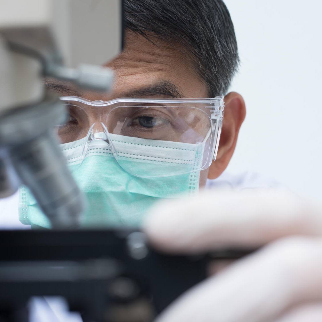
A mammogram is an X-ray image of your breasts and is the breast cancer screening technique that most people think of. It also can be used for diagnostic purposes, such as to investigate symptoms or unusual findings on another type of imaging test.
During a mammogram, your breasts are compressed between two firm surfaces to spread the breast tissue. Then an X-ray captures black-and-white images that are displayed on a computer screen and examined by a radiologist for signs of cancer. A traditional mammogram creates two-dimensional images of the breast.
At Mayo Clinic Health System, healthcare providers offer screening mammography, beginning at 40, for women at average risk of breast cancer. Average risk of breast cancer means women with no family history of breast cancer and no other risk factors.
Digital breast tomosynthesis
This screening exam, also known as 3D mammography, has become a best practice in women's health for breast cancer-detecting technology. Breast tomosynthesis differs from traditional mammograms in how the image of the breast is obtained and, more importantly, viewed.
Like a traditional mammogram, your breasts are compressed between two firm surfaces to spread the breast tissue. A digital tomographic image is created, and the radiologist can view and manipulate the images on high-resolution computer monitors that enhance visualization of the structures within the breast tissue. The tomographic images help to detect small calcifications, masses and other changes that may be signs of early cancer.
Tomosynthesis, or 3D mammograms, are best practice for all women, regardless of their cancer risk.
Watch: A demonstration of how 3D images allow breast tissue to be viewed in individual slices
Molecular breast imaging
This procedure uses a radioactive tracer and a special camera to take pictures of the breast tissue.
During the molecular breast imaging (MBI) exam, a small amount of radioactive tracer is injected into a vein in your arm. The tracer travels through your blood to your breast tissue. Cells growing quickly take up more of the tracer than slowly growing cells. Cancer cells often grow quickly and can be identified if they are taking up more of the tracer.
A special camera, called a gamma camera, detects the radiation released by the tracer. In the pictures made by the gamma camera, the cells that take up more of the tracer look brighter than the surrounding cells.
MBI provides the greatest benefit for women with dense breasts or intermediate breast cancer risk. As the density of breast tissue increases, the ability to detect breast cancer with screening mammography becomes more challenging. It's important to know that MBI does not replace, but is used in addition to, screening mammography for early breast cancer detection.
Breast MRI
Magnetic resonance imaging of the breast, also called breast MRI, is used to evaluate breast cancer or to help healthcare teams determine the causes of other problems in the breast. Breast MRI also is recommended as a screening exam for women with an elevated lifetime risk of breast cancer, such as a strong family history of breast cancer, genetic risk or other factors.
A breast MRI uses powerful magnets, radiofrequency detectors and a computer to create high-detail images of the breasts. A breast MRI may be done after a biopsy shows cancer, as a screening exam or possibly to further investigate a breast finding. Breast MRI also can evaluate the extent of any cancer, as well as evaluate the other breast.
Breast MRI may be used with mammography as a screening tool for detecting breast cancer in some people. This includes people with a high risk of breast cancer. It also includes people with a strong family history of breast cancer or who have breast cancer gene changes passed down through the family, called an inherited gene.
Talk with your healthcare team about your risk for breast cancer and the benefits, risks and limitations of each screening option. In addition, discuss all changes to your breasts you notice and ask about an appropriate screening schedule for you.
Next steps:
- Get answers to mammogram FAQ.
- Learn about the types of benign breast disease.
- Read about dense breast tissue.
- Schedule a mammogram or other breast cancer screening.
Cameron Leitch, M.D., is a radiologist in Eau Claire, Wisconsin.
This article first appeared on the Mayo Clinic Health System blog.