-
Featured News
Helping Others Heal: 3-D models improve patient care and surgical outcomes
Share this:


The soft glow of Amy Alexander's alarm clock in the darkness toyed with her attempt to fall back to sleep. Her thoughts wandered to the job she left on the printer the night before. Knowing she wouldn't rest until she knew it was running smoothly, Amy headed into work early.
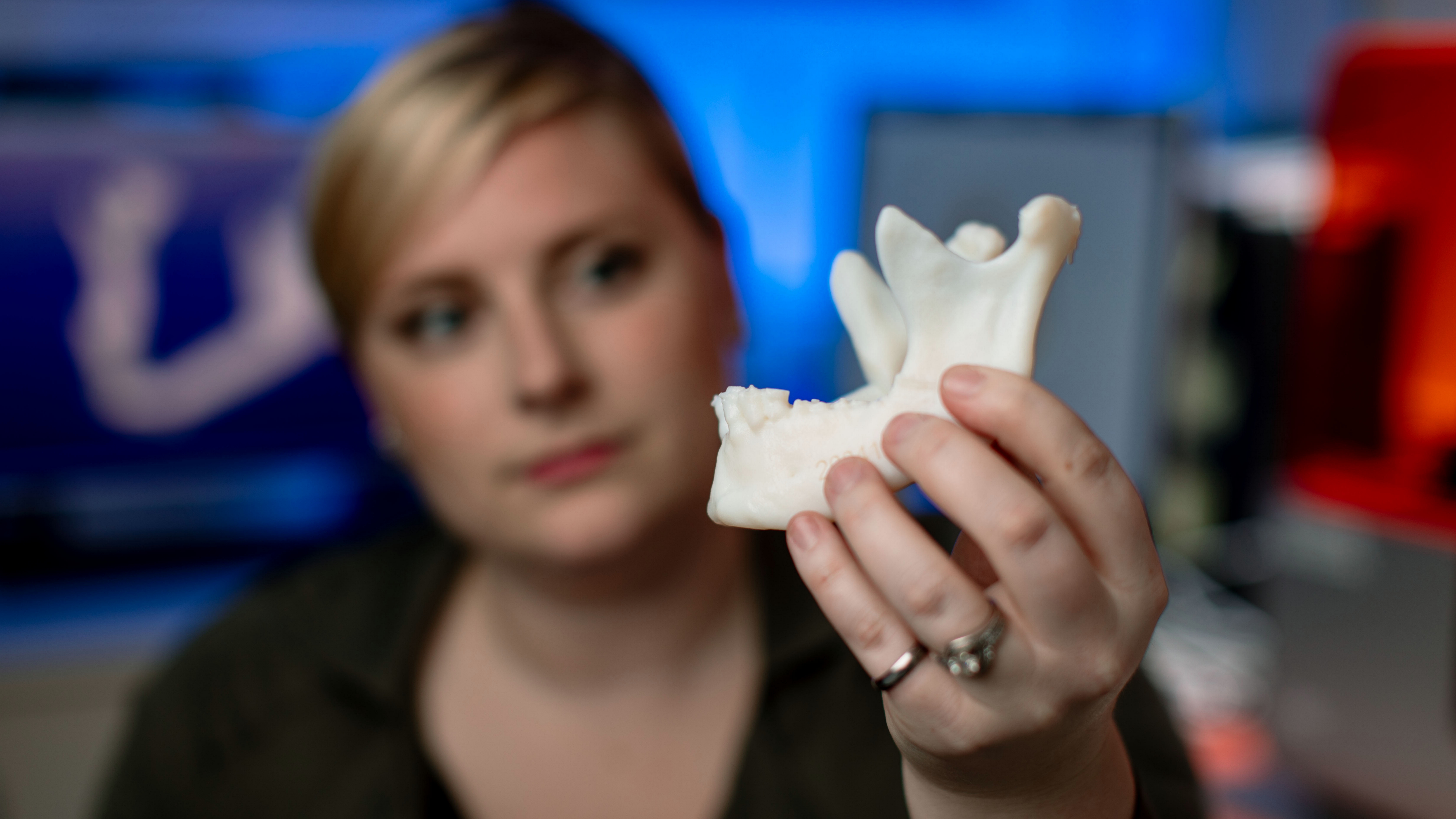
As part of the Anatomic Modeling Lab at Mayo Clinic, Amy, a biomedical engineer, pressed print the day before on a life-sized 3-D model of a 35-year-old man's face for a surgeon preparing to repair a misaligned jaw.
The model, which took about 12 hours to complete, ran through the night to be ready in time. It held the key to the surgeon's preparation to restore the man's ability to perform seemingly simple tasks such as eating solid foods.
For this particular craniofacial surgery, the surgeon planned to take three pieces of the patient's leg bone and attach it to the jaw — a procedure he had done many times before. But, because he had a model of the patient's exact facial anatomy, he was able to rehearse the surgery and knew in advance precisely where to make the cuts and what to expect during the operation, making the procedure quicker and less invasive. This surgical simulation supports better outcomes and faster recovery times.
"Medicine is very visual, and 3-D models represent another way to look inside a patient, look at the disease," says Jonathan M. Morris, M.D., a neuroradiologist and co-director of Mayo Clinic's 3-D modeling lab. "Surgeons can hold, manipulate and see a specific patient's anatomy with a clarity that cannot be replicated in two dimensions on a computer." Read the rest of the article.


