-
COVID-19
Radiology technologists use slow times to train to help with department’s AI projects
Share this:


Reduced patient volumes this spring gave Mayo Clinic Radiology technologists time to train and help move the department's artificial intelligence projects forward.
As the COVID-19 pandemic has upended business as usual at Mayo Clinic and globally, the Department of Radiology at Mayo Clinic in Rochester has used the opportunity to expedite a planned initiative to support Mayo Clinic's digital strategy.
Because of a reduced demand for their services as Mayo deferred non-emergency services, breast imaging, CT and MR technologists received training to help in the development of artificial intelligence algorithms.
"We are fortunate to have staff who are eager to learn new skills and take on new challenges," says Matthew Callstrom, M.D., Ph.D., chair of the Department of Radiology in Rochester. "The current situation has allowed us to move these projects forward."
The technologists' help will allow these algorithms to be brought into the clinical practice sooner than they otherwise would have been available.
Unexpected downtime and opportunity
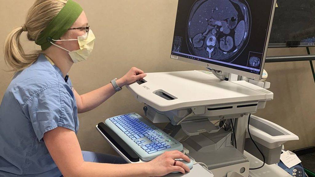
With non-emergency care deferred, there were fewer patients at Mayo Clinic, which provided CT, MR and breast imaging technologists with unexpected time to learn new skills, train and support staff in other endeavors. Many took advantage of this time to learn pancreas segmentation, which produces images that are used to teach an algorithm to detect pancreatic cancer.
In addition, breast imaging technologists trained do the same thing with breast tissue segmentation, which produces images to teach an algorithm to detect breast cancer. Another group of technologist is doing the same with liver segmentation to detect lesions.
As of this writing, nearly 2,800 cases have been completed, and more than 70 technologists have been trained. The success of the program in Rochester has allowed the program to expand to Mayo Clinic in Arizona, as well, where the group is working with Akira Kawashima, M.D., Ph.D., Abdominal Imaging.
Panos Korfiatis, Ph.D., Radiology AI Informatics, who is involved in this and other artificial intelligence projects, says more than 100 technologists will be trained to help in segmenting thousands of images. Staff who receive this training will also be able to assist on other projects in the future, with the hope that additional technologists will be given a similar opportunity.
The training program began in March with a virtual session that walked a small group of technologists through everything that is being asked of them, including lists of scans that would need to be examined and the software they would be using, Dr. Korfiatis says.
The human intelligence and effort behind artificial intelligence
"AI has received much attention, and too frequently it is viewed as magic that will solve all problems and replace us all. These projects are great examples that we still need lots of human intelligence and effort to train the AI to do tasks that we do," says Bradley Erickson, M.D., Ph.D., scientific director of the Enterprise Radiology Artificial Intelligence Subcommittee. "But if we can create large training sets such as these, it will enable us to use computers to perform many important tasks so that we can provide the best care for our patients."
Ajit Goenka, M.D., credits his team, Ananya Panda, M.B.B.S., Ishan Garg, M.B.B.S., and Garima Suman, M.D., with rising to the challenge to develop training materials and work with allied health staff in early-morning sessions. "We now will perform quality assurance and quality control processes for all segmentations, improve the training materials and then share our efforts with other institutions through a journal publication," he says.
A learning curve
Katlyn Kay, a CT technologist who is one of the early adopters helping with the pancreas image segmentation and now liver segmentation. She says the training was led by Dr. Korfiatis and his colleagues by teleconference and screen-sharing so trainees could see how they use QREADS, the system used to view clinical patient images, to outline the pancreas gave a really great idea of what was expected during this process.
She and fellow technologists who have completed the training perform the segmentations between patients. She says there is a bit of a learning curve at first.
"I like that the work helps me review my anatomy. We were given information about the anatomy around the pancreas during the training, so being able to translate that to a CT scan is a nice refresher," Kay says.
###
About Mayo Clinic
Mayo Clinic is a nonprofit organization committed to clinical practice, education and research, providing expert, comprehensive care to everyone who needs healing. Learn more about Mayo Clinic. Visit the Mayo Clinic News Network.