When Dr. Bobby Mukkamala found himself on the other side of the exam table, he relied on the cutting-edge surgical techniques at Mayo Clinic to get him back to his professional work.
While presenting at a professional meeting, Dr. Bobby Mukkamala, normally an eloquent speaker, began speaking incoherently for about 90 seconds.
"Given my age of 53 at the time, I thought it was a 'senior moment,'" says Dr. Mukkamala, an otolaryngologist and head and neck surgeon from Flint, Michigan.
His colleagues suspected he was having a stroke and convinced Dr. Mukkamala to go to a nearby emergency department for evaluation. Doctors suggested he may have had a transient ischemic attack, or ministroke. They recommended an MRI when he returned home.
That scan revealed something far more serious: a brain tumor. His journey as a patient had begun — and it would ultimately lead him to Mayo Clinic.
Finding the right brain cancer care
After sharing the news with his family, Dr. Mukkamala tapped into his professional network. "Within a week of my diagnosis, I had half a dozen Zoom calls with neurosurgeons around the country," he says. "They were all wonderful with similar but slightly different perspectives on how to approach my case."

One call, however, stood out — his conversation with Dr. Ian Parney, (pictured here) a neurosurgeon at Mayo Clinic in Rochester, Minnesota and member of Mayo Clinic Comprehensive Cancer Center.
Dr. Parney knew the tumor was large, complex and near critical speech areas in the brain. "It was important to Dr. Mukkamala to protect those areas," says Dr. Parney.
Unlike other surgeons who recommended two brain surgeries, Dr. Parney recommended a single awake craniotomy with speech mapping. During the procedure, the patient answers questions, and brain activity is monitored. This helps surgeons avoid damaging parts of the brain responsible for speech. His extensive experience — about 200 similar brain tumor procedures per year — gave hope to Dr. Mukkamala that the single operation was the best choice.
"Dr. Parney spent time answering every question we had," Dr. Mukkamala says. "That is what healthcare should be. As soon as we got off the call, my wife and kids said, 'That's it. That's where you're going.'"
Using advanced surgical techniques to guide care
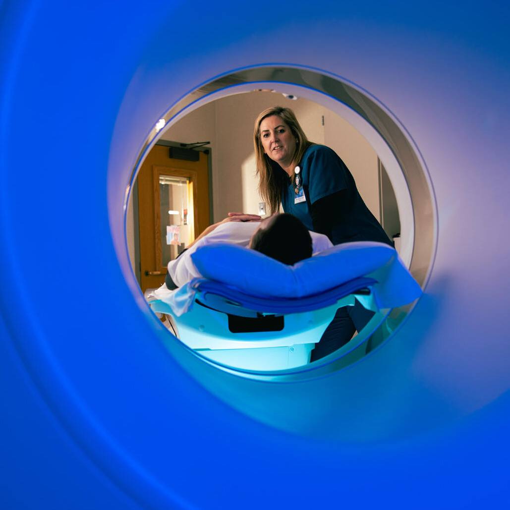
In December 2024, Dr. Mukkamala underwent an awake craniotomy with speech mapping. The surgical team also used an intraoperative MRI. This advanced imaging technique provides real-time, high-resolution MRI scans while the surgery is in progress.
"We do an MRI during the procedure to get the most accurate image so that we can remove the tumor safely," says Dr. Parney. Integrating functional imaging into image-guided systems in the operating room is a technique that Dr. Parney's team develops and tests to improve patient safety. He also correlates these techniques with novel strategies such as intraoperative electrophysiological mapping (using electrodes or electrical simulation to identify and preserve functions) and fluorescence-guided resection.
In Dr. Mukkamala's case, as part of the speech mapping, Dr. Nuri Ince, a professor of neurosurgery and biomedical engineering at Mayo Clinic, provided a novel electrocorticography technique that showed critical areas of function without requiring direct cortical stimulation (electrical signals to the brain's outer layer), as is usually necessary.

Dr. Parney and his colleagues were able to remove more than 90% of Dr. Mukkamala's tumor without damaging the speech areas. Six weeks after surgery, he was once again speaking professionally and confidently to large groups.
Coordinating multidisciplinary cancer care
Dr. Mukkamala's cancerous brain tumor was a low-grade IDH-mutant astrocytoma. This type of brain tumor arises from astrocytes (a type of glial cell in the brain) and carries a mutation in the IDH (isocitrate dehydrogenase) gene.
After surgery, Dr. Mukkamala met Dr. Ugur Sener, a neuro-oncologist at Mayo Clinic, who prescribed a new targeted drug to treat any remaining cancerous cells. The less toxic therapy allowed Dr. Mukkamala to avoid chemotherapy and radiation, which are standard treatments for brain cancer that can cause side effects such as fatigue and nausea.
"We've built one of the largest brain tumor practices in the world here at Mayo," Dr. Parney says. "We have the right resources and the right teams in place to provide cutting-edge therapies and holistic care."
Bringing new 'tumor wisdom' to the bedside
While his life today looks much like it did before his diagnosis, Dr. Mukkamala says his perspective is forever changed by his experience. "I used to be more science than emotion, but I've learned there's room for both," he says.
Dr. Mukkamala was alone when he received the news that he had cancer, much like most of his patients were when he delivered hard news. "It never occurred to me before that it was a problem to share a diagnosis when a patient was alone," Dr. Mukkamala says. He now tries to ensure his patients have support.
It's one of the many lessons he attributes to "tumor wisdom." "My brain may be a little smaller," says Dr. Mukkamala, "but I think it's happier and wiser."


