-
Featured News
Mayo Clinic 3-D models bring patient anatomy from 2-D screens to surgeons’ hands

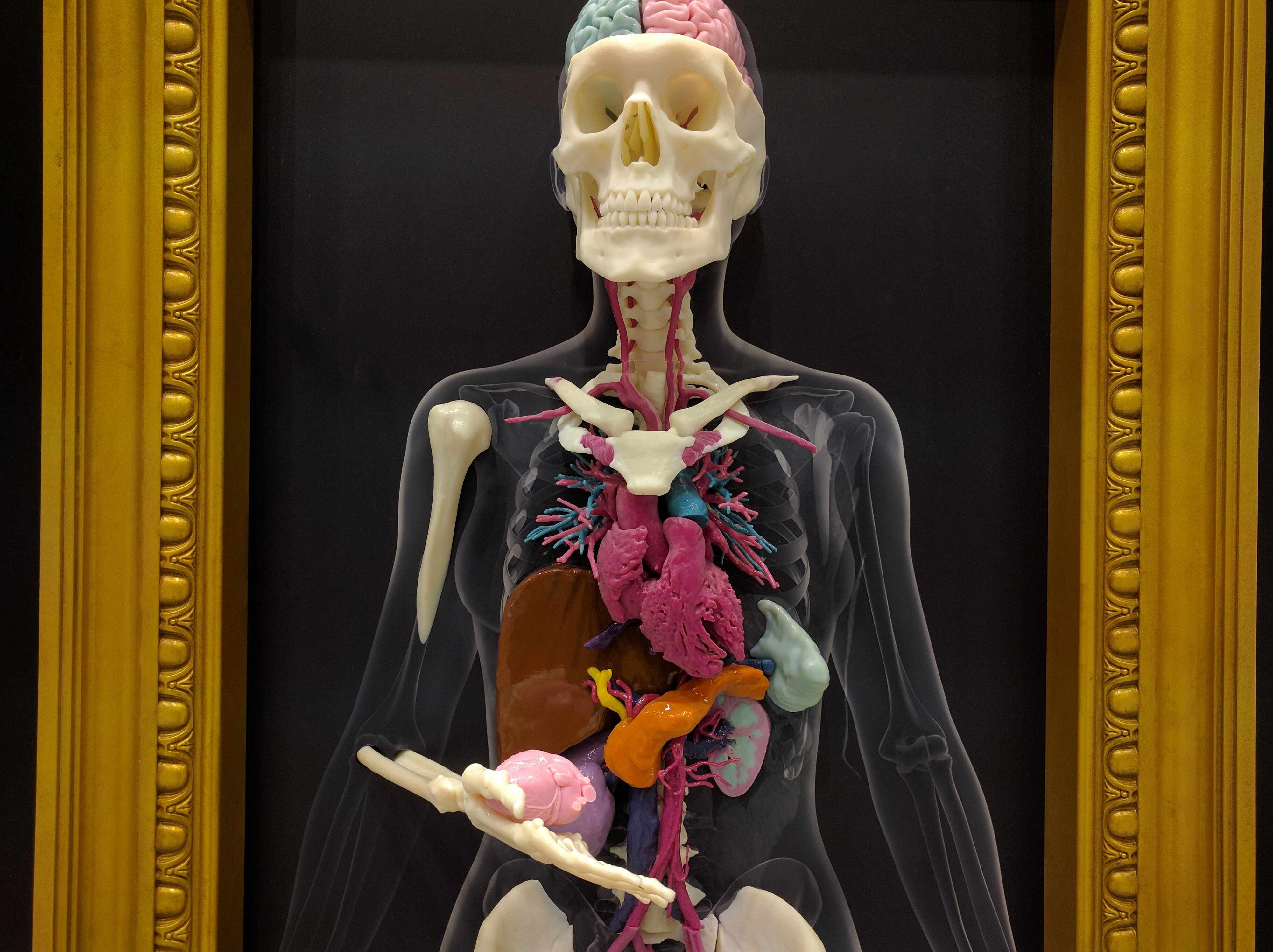
ROCHESTER, Minn. — Mayo Clinic’s 3-D anatomic modeling program started with a realization that surgeons needed a new way to look at human anatomy that went beyond two-dimensional images.
Surgeons who were planning the separation of conjoined twins in 2008 approached the Department of Radiology about producing a 3-D model of the babies’ shared liver.
The rest is history, with the 3-D anatomical modeling program growing exponentially over the past eight years. Since that time, Mayo Clinic has invested in four industrial 3-D printers in the Department of Engineering and three printers of various technologies in the Department of Radiology. They routinely run nonstop in the Department of Radiology.
“Our surgeons were really the ones to request a physical model,” says Jonathan (Jay) Morris, M.D., a Mayo Clinic neuroradiologist. The models provide a patient-specific, life-size sense of scale, helping surgeons better visualize difficult anatomy, especially when something like a tumor is changing the anatomy. Dr. Morris says that, on the screen, adult anatomy and child anatomy look the same size, for example.
“A lot of perception comes through touch, although you’re not aware of it, because we live in the physical world,” says Dr. Morris. “We work with 3-D objects all day, so you’re brain doesn’t have to come up with — how big is something, what size is that — from a collection of axial images. The same thing happens in surgery. Surgeons can stop looking at the radiographic images and trying to compile what this image looks like in real space, and how they’re going to operate on it.”
Because the 3-D models are life-size and patient-specific, surgeons are able to hold and rotate the model to get a better sense of how they will need to position the patient on the table, where they can make cuts, or whether they can make different cuts that they hadn’t thought were possible, Dr. Morris says.
MEDIA CONTACT: Ethan Grove, Mayo Clinic Public Affairs, 507-284-5005, newsbureau@mayo.edu
Watch: Dr. Morris discusses the 3-D Modeling Lab.
The models also help patients and their families better understand what is going on with their bodies and what Mayo Clinic specialists are going to do to help.
“If you give the patient a model, it allows them to physically hold their own bones, for example, outside of their body, but also allows them to understand why a surgeon might have to cut somewhere or what the risks are [and] what the benefits are,” Dr. Morris says. “What we’ve been able to do is take patients’ anatomy [off of a 2-D screen] and bring it back into the real world.”
With demand growing, Dr. Morris and his co-director, Jane Matsumoto, M.D., a pediatric radiologist, continue to work in a multidisciplinary fashion with all of the surgical and medical specialties throughout Mayo Clinic. “We’re developing new ways to take out tumors through less invasive means so that the larger surgeries don’t have to be done,” he says. “At our institution, 3-D printing is changing the landscape of oncologic surgeries, simulation, patient education, and device creation. It is bringing in-house manufacturing into the hospital setting in a way never possible before. “
Drs. Morris and Matsumoto will have a display at the Radiological Society of North America’s Scientific Assembly and Annual Meeting in Chicago. They also are co-founding the 3-D Printing Special Interest Group, a collaborative community for Radiological Society of North America members. The co-directors will give demonstrations, present research and share their expertise with radiologists from around the Western Hemisphere at the conference, which runs through Friday, Dec. 2.

###
About Mayo Clinic
Mayo Clinic is a nonprofit organization committed to clinical practice, education and research, providing expert, whole-person care to everyone who needs healing. For more information, visit http://www.mayoclinic.org/about-mayo-clinic or https://newsnetwork.mayoclinic.org/.
Related Articles


