Share this:


Mayo Clinic recently became the first to use a next-generation 4D intracardiac echo (ICE) device in patients to guide heart procedures such as left atrial appendage closure, mitral valve repair and treatment of tricuspid valve regurgitation. The technology can also help physicians perform procedures without putting patients under general anesthesia.
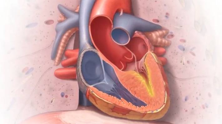
An echocardiogram is a noninvasive test that uses sound waves to produce detailed images of the heart's size, structure and function, as well as detailed images of the heart's valves. An echocardiogram also can be used to measure the heart's blood volume, and the speed and direction of blood flow through the heart.
Echocardiograms can be performed in several ways, including on the surface of the chest, or transthoracic echo, and via the esophagus, or transesophageal echo. But 4D echo takes things further, providing a detailed view captured from inside of the heart.
"The next generation 4D ICE technology can transform how we perform some complex transcatheter interventions. For example, it facilitates the performance of left atrial appendage closure under moderate sedation, making the procedure accessible to many patients who are not good candidates for general anesthesia. It also provides excellent imaging of the tricuspid valve, allowing of a more effective transcatheter treatment of tricuspid regurgitation," says Dr. Mohamad Adnan Alkhouli, an interventional cardiologist at Mayo Clinic in Rochester.
Watch: Dr. Mohamad Alkhouli discusses the new technology
Journalists: Broadcast-quality sound bites with Dr. Alkhouli are available in the downloads at the end of the post. Please courtesy: "Mohamad Alkhouli, M.D. / Cardiovascular Disease / Mayo Clinic."
The new 4D echo catheter, which is small enough to be inserted into the heart chambers, uses advanced imaging technology to create high-quality 3D moving images. This technology allows physicians to see structures and blood flow in live motion inside the heart, which is a major improvement over the standard intracardiac echo catheters that are only capable of providing 2D images.
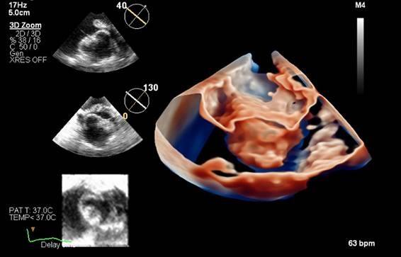
This in-motion view from inside the heart provides useful information for clinicians, especially in areas of the heart that are difficult to adequately view via transthoracic or transesophageal echo. The 4D technology provides greater visual details to guide interventional heart procedures, such as left atrial appendage closure, mitral valve repair and treatment of tricuspid valve regurgitation. Also, the 4D echo does not require the patient to be under anesthesia, which is important for patients with sedation sensitivities.
In June, Dr. Alkhouli performed the first series of in-person transcatheter interventions at Mayo using 4D ICE imaging and is working to create standardized imaging protocols for ICE-guided structural heart procedures.
"Mayo Clinic continues to be on the forefront of novel technologies that expand access to care for our growing population of patients with structural heart diseases," says Dr. Alkhouli.
Media contact:
- Terri Malloy, Mayo Clinic Public Affairs, newsbureau@mayo.edu
____________________________________________
For the safety of its patients, staff and visitors, Mayo Clinic has strict masking policies in place. Anyone shown without a mask was either recorded prior to COVID-19 or recorded in a nonpatient care area where social distancing and other safety protocols were followed.
Related Articles