-
Cancer
Mayo Clinic Minute: Lighting up lung nodules during surgery
Share this:





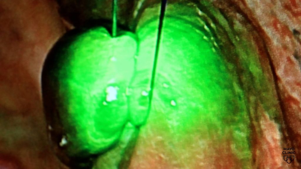
A tiny lung nodule can be difficult to find during surgery. Now, Mayo Clinic surgeons are using fluorescent imaging technology to help locate suspicious nodules with greater precision while preserving healthy lung tissue.
The imaging contrast substance is administered before surgery and travels through the body, binding to certain lung cancer cells. During the procedure, surgeons use a specialized camera to detect the fluorescent signal, causing the cancerous tissue to glow. This allows the surgical team to easily identify and remove targeted tissue.
Journalists: Broadcast-quality video (1:06) is in the downloads at the end of this post. Please courtesy: "Mayo Clinic News Network." Read the script.
"Using this fluorescent agent is really helpful because it hones to the cancer cells and binds to one of the receptors on cancer cells," says Dr. Jonathan D'Cunha, a Mayo Clinic cardiothoracic surgeon. "When you're in the operating room, you can use your advanced fluorescent imaging to see the tumor light up."
The technology can help surgeons identify tiny nodules that may be difficult to see or feel during minimally invasive procedures. It also helps distinguish cancerous tissue from healthy lung tissue, allowing for greater precision during surgery.
"We're always looking for tools to be more precise in what we do," says Dr. D'Cunha. "The earlier we can identify and resect, the better chances and better outcomes for patients."
Learn more: Mayo Clinic Lung Cancer Program