-
Cancer
Mayo Clinic Q and A: Breast density reporting and supplemental testing
Share this:


DEAR MAYO CLINIC: I recently had my annual screening mammogram and was told everything was normal. However, I just read an article that said health care professionals must inform patients if they have dense breasts. My clinician did not share anything. Why is this information important? How do I know if I have dense breasts? What additional screening is required?
ANSWER: Breasts come in all shapes and sizes. Breast density is not determined by how a breast looks or feels. Breast density refers to the amount of fibroglandular tissue in a woman's breast compared to fatty tissue.
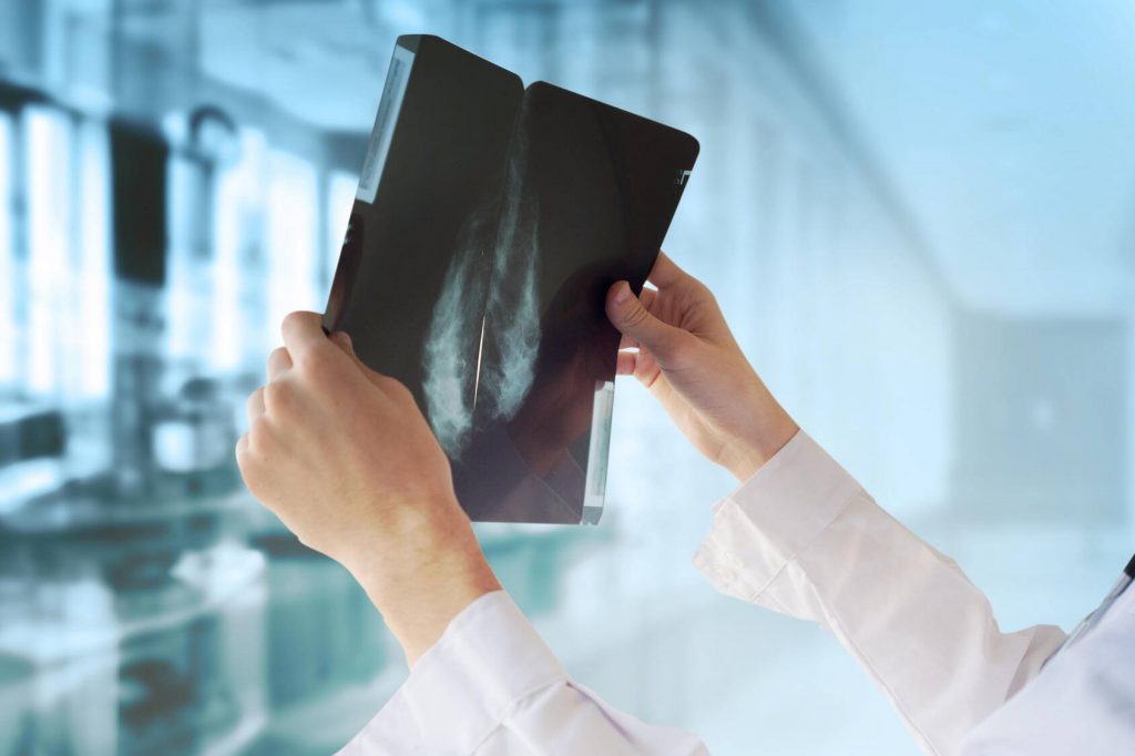
Fibrous connective tissue gives the breast shape, and glandular tissue responds to hormonal influences, produces milk and is where breast cancer forms at the cellular level. Fat in the breast is, well, just fat. On a mammogram, the fibroglandular tissue looks white, while the fat looks black. The whiter the breast appears on a mammogram, the denser it is considered.
Dense breast tissue by itself does not need to cause alarm. Approximately 40% to 50% of women have dense tissue. But women with dense breast tissue are at a higher risk for breast cancer compared to women without dense breasts. In addition, dense tissue can mask abnormalities on the mammogram, making it harder to detect small cancers.
Generally speaking, mammograms can detect approximately 80% of breast cancers in women without dense tissue, but that number decreases to below 60% in women with dense breast tissue. The more dense the breast tissue appears on a mammogram, the further the detection rate falls. That is one of the reasons for the most recent ruling by the Food and Drug Administration that aims to help further standardize information that health care organizations share. In 2009, Connecticut enacted the first breast density reporting law requiring facilities performing screening mammograms to inform patients about breast density, and this more recent ruling will standardize reporting at the federal level.
Mayo Clinic has been providing women with information about breast density for years.
Data show us that mammograms consistently reduce breast cancer mortality by detecting breast cancer at smaller sizes and earlier stages compared to women who do not have mammograms, regardless of breast density. Mammograms still detect microcalcifications and areas of abnormal tissue that might indicate cancer is hiding. But women who have dense breast tissue should discuss the option of supplemental screening in addition to undergoing a yearly mammogram.
Breast density is only one factor that contributes to someone's overall risk of developing breast cancer. The decision to undergo additional imaging may depend on your age, exactly how dense your tissue is, your personal preferences and whether you have additional risk factors for breast cancer, including family history of breast cancer or ovarian cancer, prior breast biopsies, or exposure to hormones.
Today, there are several options for breast imaging that may be used in conjunction with the mammogram for women with dense breast tissue, including:
- Tomosynthesis, or a 3D mammogram
This is performed at the same time as the traditional 2D mammogram. Tomosynthesis only increases cancer detection slightly, but it does reduce the likelihood of false positive findings. This translates to a lower recall rate — getting the dreaded phone call to return for an additional imaging. - Whole breast ultrasound
This technique has been used historically for women with dense tissue, although the added cancer detection rate is relatively low compared to using mammography alone. The major downside is that whole breast ultrasound has a fairly high false positive rate. - Contrast-enhanced digital mammography
This can be done at the same time as the traditional screening mammogram, but uses an iodine-based contrast, delivered through an IV prior to the test. This is a newer imaging strategy, but initial research studies have been very promising. - Molecular breast imaging (MBI)
MBI is a test that uses a radioactive tracer and a special camera that looks at the energy level of cells. Cells that are growing quickly take up more of the tracer than do slowly growing cells. Cancer cells often grow quickly, so the idea is that they will take up more of the tracer. MBI is a great option for women with dense tissue who do not have a lot of other risk factors for breast cancer. - MRI
This test has the highest cancer detection rate, finding significantly more cancers than mammograms alone. MRI is the costliest test to image breast tissue, and current guidelines recommend MRI only for women at the highest risk for breast cancer — women whose lifetime risk exceeds 20% to 25% based on several factors. MRI also is beneficial for women previously diagnosed with breast cancer and who have dense breast tissue, or if diagnosis occurred before age 50.
The decision to pursue any of these options should be made based on several factors, including which tests are accessible in your area, your insurance coverage, the pros and cons of each test, and your individual cancer risk.
Your primary care clinician is a good person to have conversations with, but you also can ask for a referral to a breast health specialist who can help guide you further to ensure you have the most appropriate imaging based on your breast density and overall cancer risk. — Dr. Lauren Cornell, Hematology/Oncology, and Dr. Kristin Robinson, Radiology-Diagnostic, Mayo Clinic, Jacksonville, Florida
****************************
Related Articles
- Mayo Clinic Minute: Determining if you have dense breasts published 10/13/22
- News Release: Mayo Clinic expert offers guidance on supplemental screening for women with dense breast tissue published 10/19/21