-
Research
Science Saturday: Glioma Forecast? Excellent Chance for Better Weather says Researcher
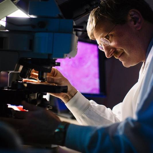
Entering the Mathematical Neuro-Oncology lab on Mayo Clinic’s location in Phoenix, Arizona, is like walking onto the bridge of a ship in some fictional universe. There's a definite "Star Trek" vibe.
"Yes, 'Star Trek' is good," laughs Kristin Swanson, Ph.D., a mathematical oncologist at Mayo Clinic, "but I like 'Big Bang Theory.' I kind of joke that that's what my lab feels like." Geeky or not, she and her team use math to study glioblastoma, a type of cancer that starts in the glial cells of the brain and spine. Normally, these cells support and protect neurons, but when they become cancerous, they divide without stopping, forming tumors that insinuate themselves into the brain. About 80% of malignant brain tumors are gliomas.
"It's what John McCain passed away from, and Teddy Kennedy and Beau Biden," she says. "It's a particularly aggressive tumor that has a median survival of about 14 months. And that rate has barely moved over the last 50 years. It's just an unbelievably challenging disease."

But by applying her imagination and grasp of math, and channeling her own sense of loss after a family member’s death from cancer, Dr. Swanson is making sense of vast data sets collected from thousands of patients with brain tumors. With artificial intelligence (AI) and other mathematical modeling methods, she's getting to know glioblastoma at a whole new level — its texture, density, direction of growth and rate of invasion. And she's making patient-specific maps of tumors to predict which treatments will work best to terminate them.
Today's Forecast
Although the standard treatment for glioblastoma is aggressive — surgery followed by chemotherapy and radiation — tumors respond in widely different ways. Only 40% of patients survive for at least one year, but almost 6% live much longer, more like 60 months.
So when it comes to predicting glioma growth, average doesn't mean much.
Cancer care teams have had meager means by which to gauge what each patient might expect. And, worse, the teams have minimal evidence to indicate if a treatment is significantly affecting tumor growth. A person's glioma may slow its growth under a drug regimen, or after surgery or radiation, but it's been impossible to figure out how significant that reduction might be. Without treatment, would the tumor have doubled in size, tripled or quadrupled? There's been no way to judge.
What's needed are tools to predict what the disease is going to do in each patient, with or without a particular treatment. The system that Dr. Swanson has built achieves this in a way that's surprisingly analogous to weather prediction.
Basically, it's a tumor forecasting center.
Whereas a hurricane center uses weather satellite data to predict storm paths, Dr. Swanson's lab uses MRI data to predict glioma growth. The result is a video showing the predicted movements of the tumor within the brain over days and months, which can be shared with the patient.

Both types of predictions rely on a branch of AI called "machine learning," which is based on the premise that computer systems can learn by example. AI is the key to driverless cars and the inner ear of speech recognition (hey, Alexa, play "Space Lab").
For Dr. Swanson, it's the forecaster of tumor behavior.
"We track with MRI, and we validate with additional biopsies," she explains. "So that's the loop. We learn on some biopsies, and we validate that our prediction is accurate on another set. And then we continually refine that using artificial intelligence tools." So far, they've collected samples from 3,000 patients and 80,000 MRIs. That's a lot of data. AI makes sense of it all, looking at the characteristics of cells in the same area of a tumor, called it’s microenvironment, and plotting the path and pace of cancer invasion.
Animated Tumor Maps and 3D-Printed Brains
In weather prediction, radar locates precipitation; calculates its motion; and estimates its type, such as rain, snow or hail. Sonar profiles wind. AI uses that radar and sonar data to determine the structure of storms and their potential to cause severe weather.
In an analogous way, using MRI images and biopsies, Dr. Swanson's lab defines glioma habitats; locates broken genes; and tracks responses to surgery, radiation and drugs. AI draws real-time 3D tumor maps, and mathematical modeling generates patient-specific animations that forecast how that tumor will grow through time.

There's an immediate advantage to mapping tumor cell invasion in 3D. Tumor cells actually invade well beyond the imaginary edge of an MRI image, and these boundary regions represent a diffuse but insidious faction of the invasion. With modeling, explains Dr. Swanson, researchers can identify where those cells are and optimize radiation dosing so the dose matches the tumor cell density. “We're designing a clinical trial for that right now, to make that as rote within the Mayo system," she says, adding that for some patients, “we've 3D-printed their brain, their specific brain, (showing) their specific lesion within it."
Embracing Diversity
Every patient is different. Every tumor is different. "We all know that," explains Dr. Swanson. "We say that, but people don't hear it. The heterogeneity (diversity in appearance of cancers under the microscope) across patients is unbelievably wide, and for the most part, it goes unexplained."
For one thing, the genetic makeup of tumors is diverse, not just within a cancer type, such as neuroblastoma, non-small cell lung cancer or acute lymphoblastic leukemia, but rather within every patient. In any given tumor, different faulty genes cause different problems in different cells. This genetic diversity makes every glioblastoma a mosaic of related yet distinct cancers, each in its own unique evolutionary landscape.

Until recently, these differences made it difficult to find effective treatments. Now they have the potential to turn up the power of personalized cancer treatment because the processes that generate and maintain the genetic diversity of tumors might provide new therapeutic targets. To explore heterogeneity, Dr. Swanson uses the MRI data to subdivide tumors into regions based on variations in cell density, fluid level, blood flow and so on. The distribution of these distinct habitats affects the course of the disease.
"In order to succeed in this disease, you need to know what's happening where, and what can I target that region with," Dr. Swanson says.
Identifying Targets
One of the ways that cancer cells differ from healthy cells is a characteristic called "growth factor independence," which means that they don't need stimulation from external growth factor signals to multiply.
Cancer cells can achieve this independence by:
- Making their own growth factors
Gliomas have this ability, and they can generate an unlimited supply of a signal called platelet-derived growth factor (PDGF). - Making too many receptors for growth factors
Gliomas can do this too. They're known to overproduce both PDGF receptors and epidermal growth factor (EGF) receptors. - Making receptors that malfunction, producing cellular responses even when the external signals aren't present
Gliomas even have this skill.
Because growth factors are clearly bad actors, Dr. Swanson's lab has been exploring their healthy and pathological roles. They've modeled the release of PDGF in brain tissue with varying dynamics. Their simulations showed that outcomes depend a lot on where the growth factor is coming from within the tumor. Is it in a poor ecological niche with little blood flow? Then you're likely to see localized, short-duration growth. Is it in a rich habitat? Then you're liable to see widespread, chronic growth. In addition, they found that the tumors amplify EFG or PDFG receptors, but rarely do tumors amplify both.
With modeling, Dr. Swanson can now show patients their own specific tumor maps and point out the areas predicted to have elevated levels of growth factors or growth factor receptors. Then she can run forward through their MRI video forecast and demonstrate what would happen, for example, if they received a receptor-targeted therapy — which regions would continue to grow and which would melt away.
Tailoring Treatment
Using serial images of tumors, Dr. Swanson has shown that it's possible to divide glioblastomas into two types based simply on how quickly they grow. One type is faster-growing, yet more treatment-responsive with increased survival. The other is counterintuitively slower-growing, yet less responsive with shorter survival. She and her team built a predictive model to identify the type using information that most clinicians already would have at their fingertips immediately following radiation therapy. Depending on which group patients fall into, they may benefit from select alternative treatments more than from standard of care.
Sex is another source of heterogeneity. Glioblastoma is diagnosed nearly twice as often in men than in women, but the standard treatment is more effective in women than in men.
Dr. Swanson has been keen to learn the whys and wherefores of these differences because tailoring treatment by gender may improve survival. Recently, she and her collaborators compared gene expression and treatment response data to identify sex-specific glioblastoma subtypes. What they found was that in men with glioblastoma, the faulty genes usually are involved in cell division; whereas, in women, they're more often involved in intercellular communication. These differences go beyond hormonal influences and appear to be intrinsic to the tumor cells themselves.
Moreover, the sex of the patient correlates not only with prognosis, but also with responses to particular treatments. Using MRI "satellite" maps to track tumors through time, Dr. Swanson and her collaborators were able to visualize cancer therapy response patterns. Under standard treatments used routinely at Mayo Clinic, male tumors tended to respond strongly but transiently; whereas, female tumors responded moderately and sustainably.
Insights like this can help pathologists distinguish female and male cancers at the molecular level, and may allow oncologists to optimize the therapeutic regimen in a patient-specific way.
Math and Cancer
Patient-specific maps of tumor invasion and the path of their destruction will become more detailed as information from patient MRIs expands Dr. Swanson's database, and as AI validates her predictive models. By its nature, the system continuously improves itself. Science without fiction.
The goal is to give patients and their cancer care teams the most accurate forecasting possible. Because "without a patient-specific prediction of what that tumor would do without treatment, we wouldn't know what the impact of the therapy really was," Dr. Swanson explains. As each person's cancer is treated, the system zooms in on the paths of tumor growth to see what's working where.
In the past, medicine progressed by focusing on the average results of a treatment in a group of seemingly similar patients. With glioblastoma, that only took things so far. Today, the efforts of Dr. Swanson and her team, as well as others, are making it possible to care individually for each person. And that fulfills the lab's motto: "Every patient deserves their own equation."
— Megan McKenzie, July 2019


