-
Seeing Disease: A Vision for Better Imaging
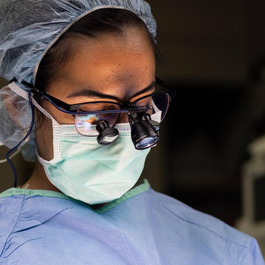
For a patient with cancer in the bones of his pelvis, Matthew Callstrom, M.D., Ph.D., a Mayo Clinic interventional radiologist, was operating using minimally invasive tools, such as thin needles and catheters to reach and destroy tumors.
In this case, he was trying to insert a hollow needle carrying supercold fluids into the tumor to freeze and kill the cancer. But the angles of the pelvis are complex, aiming is difficult, and the placement must be precise. Dr. Callstrom relied on computerized tomography (CT) scans to guide him. But a CT scan shows only a single slice of the body well, and trying to thread the needle through 3D space was like trying to gauge distance with one eye closed.

For a different angle, Dr. Callstrom wheeled up a fluoroscopic X-ray system, which can image the procedure in real time from any angle. It was available because Dr. Callstrom was working in a new hybrid procedural suite at Mayo Clinic Hospital — Rochester, Saint Marys Campus in Minnesota. Unlike nearly all operating rooms, this procedural suite is outfitted with all the ready-to-use imaging equipment that a surgeon might need, from MRI to ultrasound.
With the help of the fluoroscopic X-ray, Dr. Callstrom plotted a path to the tumor. Because CT is much better at revealing soft tissue and can even "see" the formation of ice, Dr. Callstrom switched back to CT to watch as ice covered the tumor and stop before he reached the nearby healthy tissue.
Embedded in Care
The hybrid procedural suite is one of the payoffs of a partnership between Mayo and Philips, the multinational Dutch health service and equipment company, to improve the use of technology, especially in cancer treatment. The alliance is so close that Philips has moved into office and research space in the new 89,000-square-foot One Discovery Square Building in downtown Rochester.
"Usually there's a big gap between the academic community and the practicing community," says James Pipe, Ph.D., a Mayo Clinic radiology researcher who has been working with Philips to develop higher-resolution, higher-speed MRI. "One way to bridge that gap is to work closely and put yourself in an environment where things get used."

"The reputation of Mayo is, of course, world-renowned," says Joland Rutgers, head of global research and development for MRI at Philips. "What we've experienced is that when you get together and you build a partnership, a common vision is very important. Very often, we rally around common visions."
Building the Suite
Dr. Callstrom's vision was having all the important imaging technologies in the operating room. They also had to be able to link and communicate among platforms so surgeons could use them in succession or even simultaneously. Without this, surgeons often would close up a patient and wheel the patient into another unit for an MRI to check that a tumor was completely removed. "It's closer to 'MacGyver,'" says Dr. Callstrom, referencing the inventive TV character who improvised solutions. "We pull things together as we can."
Dr. Callstrom began planning the hybrid procedural suite in 2015. Mayo Clinic uses imaging equipment from many large companies, including Philips, Siemens and GE. So right off, Dr. Callstrom and colleagues faced a decision: Pick the equipment from several companies and try to make it work together, or to rely on a single organization? And, if so, which one?
"We elected to go with one to try to gain a high level of endorsement so that we could integrate," says Dr. Callstrom. And they chose Philips. "One of the things that Philips brought forward was a commitment to try to drive toward solutions."
David Woodrum, M.D., Ph.D., Dr. Callstrom's colleague in Radiology, says, "We need a partner to whom we can say, hey, our clinical need is to have this. Can you bring together all your imaging platforms and create software that's compatible with all of them?"
The suite soon took shape and came online in November 2017. Additional imaging equipment was added to an adjoining room, separated by wide garage-style doors. The first patient was treated with the full complement of imaging equipment in June 2018.

The ultimate goal for the suite in years ahead is improve cancer care for patients.
"That's the vision — get the best possible outcome, have the best impact on a patient's survival for those cases," says Dr. Callstrom.
"I have to say, from having partnered with Philips, they've actually jumped in with both feet and really tried to do the right thing. They have brought a lot of energy to the partnership," says Dr. Callstrom. "I couldn't really ask for anything more from them as a partner."
Faster, Better MRI
Anyone who has endured the startling sounds and claustrophobic confines of an MRI tube can appreciate the value of spending less time in the machine. But faster acquisition of images also leads to more efficient use of expensive equipment to save money and reduce the cost of patient care.
Dr. Pipe was already a pioneer in high-speed, high-resolution MRI when he began collaborating with Philips several years ago, developing computer algorithms to gather sharper images faster while at an institute in Phoenix. When Dr. Pipe made his move to Mayo in May 2018, Philips decided to up its game even more and move into research space in the One Discovery Square Building, then still under construction.
This fast imaging, combined with the new and simpler technologies to operate the scanner that Dr. Pipe's team is designing, opens up the ways that MRI can be used to take care of patients.

"We recognized that this was a good moment to strengthen the relationship and to really bring it to the next level by working side by side with Mayo to achieve results," says Arjen Radder, global business leader for MRI at Philips.
Dr. Pipe will move his research team into the second floor of One Discovery Square in late 2019, about the same time the Philips team will move in next door. Dr. Pipe's team will be engineering computer algorithms to run existing Philips MRI scanners in subtly new patterns to gather an image of human tissues.
High-Value MRI
Although Mayo and Philips are neighbors for the first time, Philips's collaboration with Mayo researchers goes back years. Richard Ehman, M.D., a Mayo Clinic radiologist and principal investigator for Mayo's Advanced Medical Imaging Technology Laboratory, developed technology to assess the texture and stiffness of tissue. That technology was magnetic resonance elastrography, or MRE. About 10 years ago, he realized that the technology could detect a condition called liver fibrosis — scarring of the liver — and substitute for more expensive and invasive liver biopsy.

"We realized we should get it out to the rest of the world to help patients everywhere," he says.
Mayo Clinic formed the company Resoundant Inc. to commercialize the new technology and worked with Philips, as well as GE and Siemens, to produce commercial versions. Philips alone has installed more than 200 systems over the past decade.
More recently, Dr. Ehman has teamed with Philips on what he calls "high-value MRI."
"We think of MRI as a big expensive technology that we reserve for special circumstances," says Dr. Ehman. "There are many situations now in medicine where really the first test you should do is MRI. It could be used in a very low-cost way. One way to reduce the cost is instead of doing a one-hour comprehensive exam that answers many, many questions, we'll do a five-minute procedure that answers specific questions."
Dr. Ehman's lab is using a high-capability MRI scanner, provided by Philips, to develop these new types of MRI exams.
"It turned out that Philips had the vision in this area of the high-value MRI to realize that that was a great opportunity," says Dr. Ehman.
"What you often see is that when your strategic vision is aligned, you can produce outcomes-focused results, says Joland Rutgers of Philips. "That is where we are really in sync with the leadership of Mayo: visionary, knowing what to do, rallying around common goals and concepts, and working together collaboratively to stretch the boundaries of innovation."
-Greg Breining, January 2020


