-
Research
Thinking outside the box: Uncovering a novel approach to brainwave monitoring
A 3D animation of the region of the brain called the motor thalamus (shown in red and yellow) where Mayo Clinic researchers detected more precise information about brain cell activity in patients during deep brain stimulation (DBS). DBS electrodes are placed into the motor thalamus to treat movement disorders.
Mayo Clinic researchers have found a new way to more precisely detect and monitor brain cell activity during deep brain stimulation, a common treatment for movement disorders such as Parkinson's disease and tremor. This precision may help doctors adjust electrode placement and stimulation in real time, providing better, more personalized care for patients receiving the surgical procedure.
Deep brain stimulation (DBS) involves implanting electrodes in the brain that emit electrical pulses to alleviate symptoms. The electrodes remain inside the brain connected to a battery implanted near the collarbone and controlled by a remote control. While a neurologist and neurosurgeon monitor the brain waves throughout the surgery, the monitoring typically is limited to a narrow frequency range that provides a rough snapshot of brain activity.
However, Mayo Clinic researchers used more sensitive, research-grade equipment and custom algorithms to record a broader frequency range of brain cell activity that yielded higher resolution and more precise information on when and where brain cells were firing in patients during DBS surgery.
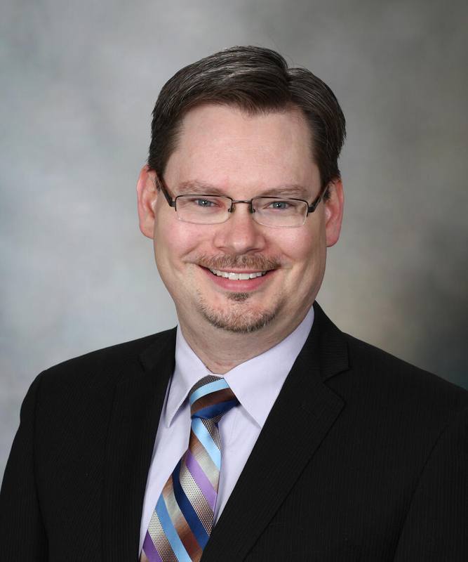
"We looked at brain activity in a different way and recorded a type of brain signal called 'broadband' that reflects the combined activity across all frequencies and is related to the firing of all brain cells in that region. We found that the broadband activity signal increased with movement and was more precise in location than the standard, more narrow frequency signal," says neurologist Bryan Klassen, M.D., lead author of the study published in the Journal of Neurophysiology.
Dr. Klassen and colleagues detected the broadband signal in the motor thalamus, a region deep within the brain that controls movement. Previous studies have detected it only on the surface of the brain.
The researchers recorded broadband signals associated with hand movement in 15 patients undergoing awake DBS. Each of the patients were instructed to open and close their hands while the researchers recorded brain cell activity in their thalamus.
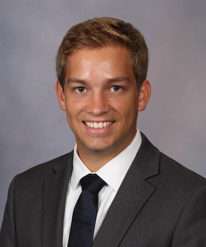
"This study enhances our understanding of how the thalamus, a brain region that is frequently targeted for deep brain stimulation, processes movement. It may lead to more precise mapping of the brain as well," says Mayo Clinic coauthor Matthew Baker, Ph.D., a postdoctoral fellow in the neurosurgery department.
Using broadband to monitor during DBS surgery may improve the treatment and outcomes for patients.
"These findings underscore the remarkable advances we can achieve through the close collaboration between the neurology and neurosurgery departments and will help us develop the next generation of brain stimulation therapies," says neurosurgeon Kai Miller, M.D., Ph.D., senior author of the study.
The next steps for this research involve further exploring how these brain activity patterns in the thalamus can be used to improve neurostimulation therapy, says Dr. Baker, a recent graduate of Mayo Clinic Graduate School of Biomedical Sciences.
"We will be investigating how this signal responds to different types of movements and whether we can use it to control new devices that only stimulate when patients need it, as opposed to constant stimulation, which is more prone to cause side effects," he says.
Review the study for a complete list of authors, disclosures and funding.


