-
Cardiovascular
Understanding your heart test: What to expect, how to prepare

Heart health is an essential part of your overall health and well-being. Your primary care provider will monitor your blood pressure and lab work during an annual physical exam or more frequently. If you have symptoms that indicate your heart may not be functioning properly, such as blood pressure higher than the recommended level, chest pain or swelling in your feet or legs, you may be referred to a cardiologist.
A cardiologist uses diagnostic testing to evaluate the heart's ability to pump and move blood and oxygen throughout your body. Hearing you need a test on your heart may be confusing or scary, so understanding the purpose of the tests can help put you at ease.
Learn about various tests to help diagnose heart diseases and conditions.
What's a cardiac stress test?
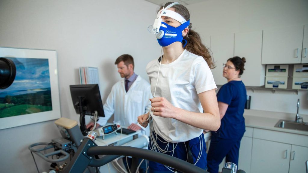
A cardiac stress test shows how your heart works during physical activity. Also known as an exercise stress test, it can detect problems with blood flow when the heart is pumping faster and harder than usual. The test can be done while walking on a treadmill or riding a stationary bike. Or you may be given a drug to mimic the effects of exercise while in a seated, resting position.
You may have a stress test to:
- Diagnose if coronary artery disease, which is the buildup of cholesterol deposits or plaques, is causing damage or disease in your coronary arteries.
- Identify arrhythmias that cause your heart to beat too fast, slow, or irregularly.
- Determine when to proceed with cardiac surgery or valve replacement treatments.
A nuclear stress test may be recommended if an exercise stress test doesn't reveal the cause of your symptoms. During a nuclear stress test, short-lived radioactive medications are injected into the body and taken up by the heart to create an image of the heart's function at rest and with stress. This medication rapidly leaves the body after the test and is safe and painless.
What's a coronary CT angiogram?
During a coronary CT angiogram, advanced CT technology and injected dye are used to take high-resolution 3D images of your heart. The 3D pictures of the beating heart and major blood vessels are used to identify coronary artery blockages. From the 3D images, your healthcare team can see if there are plaque or calcium deposits in the arteries.
What's an echocardiogram?
Sound waves produce images of your heart during an echocardiogram. This is a common test to see your heart beating and pumping blood. The images from an echocardiogram can help identify heart disease, detect congenital heart defects before birth and check for problems with the valves or chambers of your heart.
You may have one of several types of echocardiograms:
- Transthoracic
The standard type of echocardiogram that converts sound wave echoes into moving images. - Transesophageal
Provides more detailed images than the standard echocardiogram. - Doppler
Can be used with transthoracic or transesophageal echocardiogram to measure the speed and direction of blood flow in your heart. - Stress
Ultrasound images of your heart before and after physical activity can be used to check for coronary artery problems.
What's an electrocardiogram?
An electrocardiogram (EKG or ECG) is a noninvasive and painless way to quickly detect heart problems and monitor the heart health of people of all ages. You may have an ECG if you have symptoms such as chest pain, dizziness, confusion, heart palpitations or shortness of breath.
Small, adhesive electrodes are attached to your chest, arms and legs during an ECG. The electrodes record the electrical activity of your heart, and the ECG machine prints out a chart of your heart's electrical signals.
An ECG can be used to diagnose many common heart problems and may detect:
- Arrhythmias or abnormal heart rhythm
- Blocked or narrowed arteries in your heart or coronary artery disease
- If you have had a previous heart attack
Continuous ECG monitoring may be recommended if your symptoms come and go, which causes difficulty with a standard ECG test.
The two types of remote or continuous monitors are:
- Holter
A small device you can wear records 24 to 48 hours of continuous ECG monitoring. - Event
A portable device that records heart electrical signals for a few minutes at a time, typically over the course of 30 days.
What you can do to prepare for a heart test
Preparing for diagnostic heart tests helps ensure the results are accurate. Talk with your healthcare team about your questions when preparing for the test recommended specifically for your symptoms.
Here are a few general steps you can take before a heart test:
- Notify your care team of prescription medications, over-the-counter drugs, supplements or herbal products you are taking. Your heart can be affected by some medicines. Your healthcare team will tell you if adjustments are needed to your medications for the test.
- You may need to fast before specific tests. This means you may have to avoid food or beverages before the test. Your healthcare team will give you detailed fasting instructions if necessary.
- Wear loose, comfortable clothing that allows access to your chest, arms and legs. You may need to wear a hospital gown during the test. Avoid applying lotion, oil or powder to your skin before specific tests, like an ECG.
- Limit strenuous activity that may elevate your heart rate for a few hours before the test. Consuming caffeine or nicotine before some tests can affect your heart rate and interfere with the results.
- Some tests require sedation or other medicine to help you relax. This can affect your ability to drive after the test, so you need to arrange a ride to and from the healthcare facility.
- Follow specific instructions from your healthcare team.
Michael Meyers, M.D., is a cardiologist in La Crosse and Sparta, Wisconsin.
This article originally posted to the Mayo Clinic Health System blog.


