Share this:


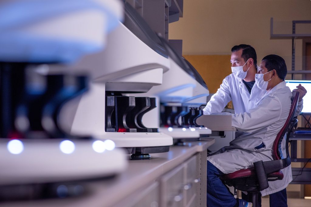
In January, as part of Mayo Clinic’s Digital Pathology Program, the Division of Anatomic Pathology in Mayo’s Department of Laboratory Medicine and Pathology in Rochester, Minnesota, transitioned a significant portion of its clinical practice, including both surgical and biopsy cases, into digital workflows.
The transition means all surgical — or frozen section — biopsy slides will be scanned into digital images prior to a pathologist’s review. Historically, patient tissue has been evaluated by generating glass slides, applying stains, and evaluating them with a microscope. Scanning slides digitally allows pathologists to take action that previously required a series of physical handoffs within analog workflows before it could be completed.
While converting glass slides into digital images means adding about one hour to each case, Joaquin Garcia, M.D., medical director of the Digital Pathology Program, believes the overarching gains are worth that investment. “In exchange for time, we get a digital format that we can share frictionlessly between medical secretaries, residents and fellows, and multiple consultants,” he says.
“Also, if a consultant needs a specialty opinion, he or she can seek that from another colleague down the hall, or by leveraging digital image access to Mayo Clinic consultants in Arizona, Florida, Rochester, and the Mayo Clinic Health System. Once the slides are scanned, we can share these digital images and really bring exponential expertise to that case within seconds.”
Read the rest of the article on the Mayo Clinic Laboratories blog.
____________________________________________
Other Mayo Clinic medical research websites:
- Research at Mayo Clinic
- Discovery’s Edge
- Advancing the Science
- Forefront
- Center for Individualized Medicine
- Center for Regenerative Medicine
- Center for the Science of Health Care Delivery
- Mayo Clinic Cancer Center