-
Featured News
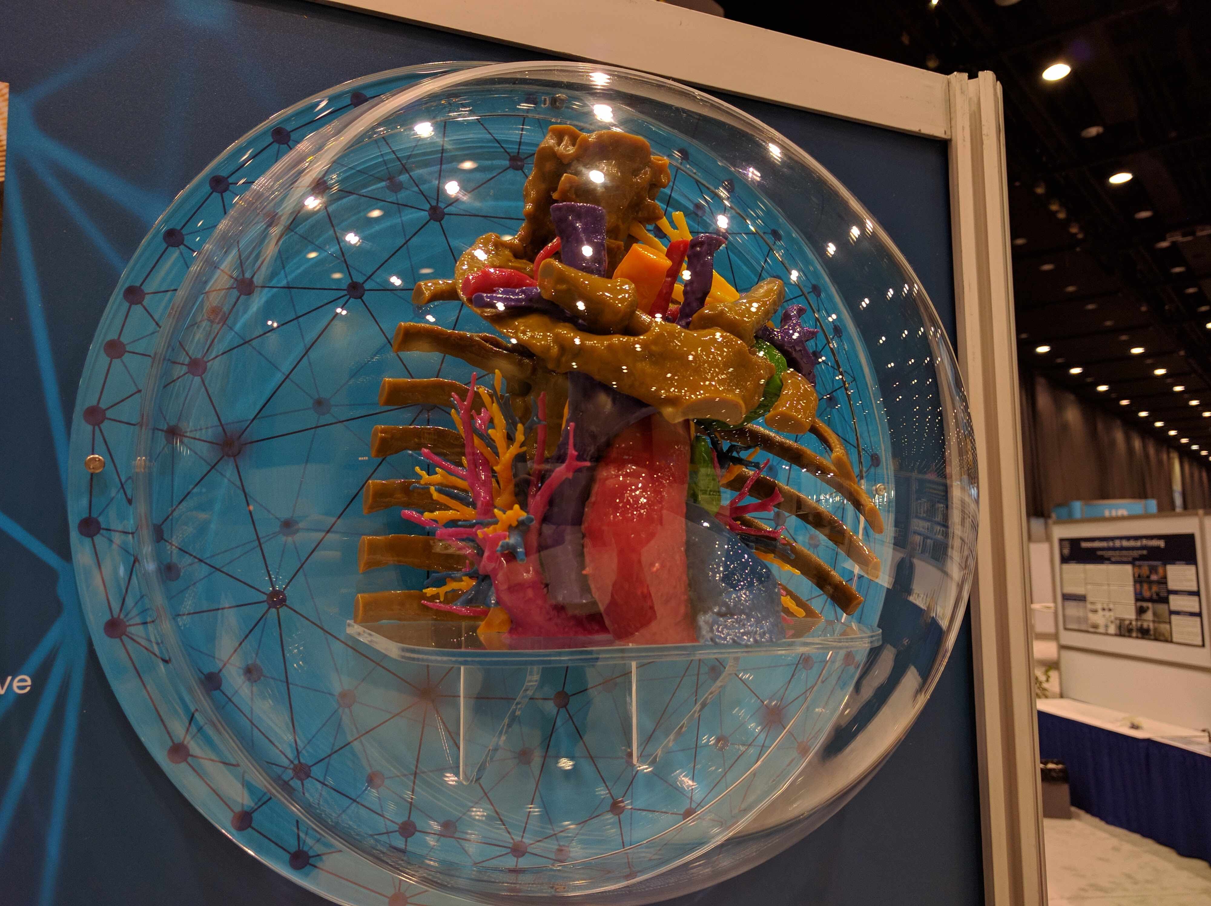
3-D Imaging in Medicine display shows anatomy of 3-D modeling program at Mayo Clinic
ROCHESTER, Minn. — Mayo Clinic’s 3-D anatomic modeling program continues to expand — stretching across multiple rooms in Mayo Clinic Hospital — Rochester, Saint Marys Campus, and down to Mayo Clinic’s Arizona and Florida campuses. And for 10 days, it also will reach into downtown Rochester.
From June 12-28, a display in Hage Atrium in the subway level of the Siebens Building will feature the history and geographical and departmental reach of 3-D anatomic modeling at Mayo Clinic. The public can view the display weekdays from 8 a.m. to 5:30 p.m. CDT.
The display puts into perspective the years of work that have touched nearly every medical specialty at Mayo.
“We know how many models the lab has produced, but it’s hard to imagine the breadth of it until you see the sampling we have here in this display,” says Jane Matsumoto, M.D., co-director of the 3-D modeling lab in Rochester. “In getting this historical perspective of the practice, you start to see the evolution. It’s really fascinating to see the first models we did and compare them with the models we are doing now.”
None of this would be possible, though, were it not for the medical need for personalized patient care, and the enthusiasm of the physicians and surgeons who request the models, says Jonathan Morris, M.D., who also is co-director of the Rochester lab. “The first model we did was requested by the surgeons who were going to be separating conjoined twins. They needed this next-level visualization of the patients’ anatomy to better see and plan out the complex surgery.”
The models have been — and are — used in diagnoses, surgical planning and prep, and in some cases a rehearsal before a surgery. Each is unique, representing a specific patient’s anatomy. After resection, surface scanning technology employed in anatomic pathology can be used to capture a 3-D image of the specimen to help understand the surgical margins, best dissection technique, and in medical education. These surface scanned models reproduce a specimen so vividly that they are unparalleled educational tools for patients and healthcare providers alike.
“Medicine is very visual, and 3-D models represent another way to look inside a patient, look at disease,” Dr. Morris says. “It takes a 3-D image on a two-dimensional surface and makes it real. Surgeons can hold, manipulate and see a specific patient’s anatomy with a clarity that cannot be replicated in two dimensions on a computer. This, in turn, shows them the path the surgery should take.”
The demand for 3-D models has led to an expansion of the program to Mayo’s Arizona and Florida campuses, with Rahmi Oklu, M.D., Ph.D., and Robert Pooley, Ph.D., respectively, leading the new labs.
“We are excited about our 3-D printing services here in Florida and hope to continue to expand capability,” Dr. Pooley says. The Departments of Laboratory Medicine and Pathology and Anatomy are working hard to incorporate 3-D scanning and printing into the medical school curriculum. The technology offers the possibility to replicate rare and unusual specimens only available at the Rochester campus and make the available to students at other sites.
Dr. Oklu says his Arizona lab has produced more than 50 models. “I’m excited about the work we’ve done so far, and looking forward to taking our program to the next level,” he says.
“With these patient-specific models, we are able to provide integrated, high-quality care to patients with tumors, fractures, malformations and more,” Dr. Matsumoto says. “This program really brings people together across specialties. It shows there are no boundaries.”
###
About Mayo Clinic
Mayo Clinic is a nonprofit organization committed to clinical practice, education and research, providing expert, whole-person care to everyone who needs healing. For more information, visit mayoclinic.org/about-mayo-clinic or newsnetwork.mayoclinic.org.
MEDIA CONTACT
Ethan Grove, Mayo Clinic Public Affairs, 507-284-5005, newsbureau@mayo.edu


