Cancer
October 10, 2024
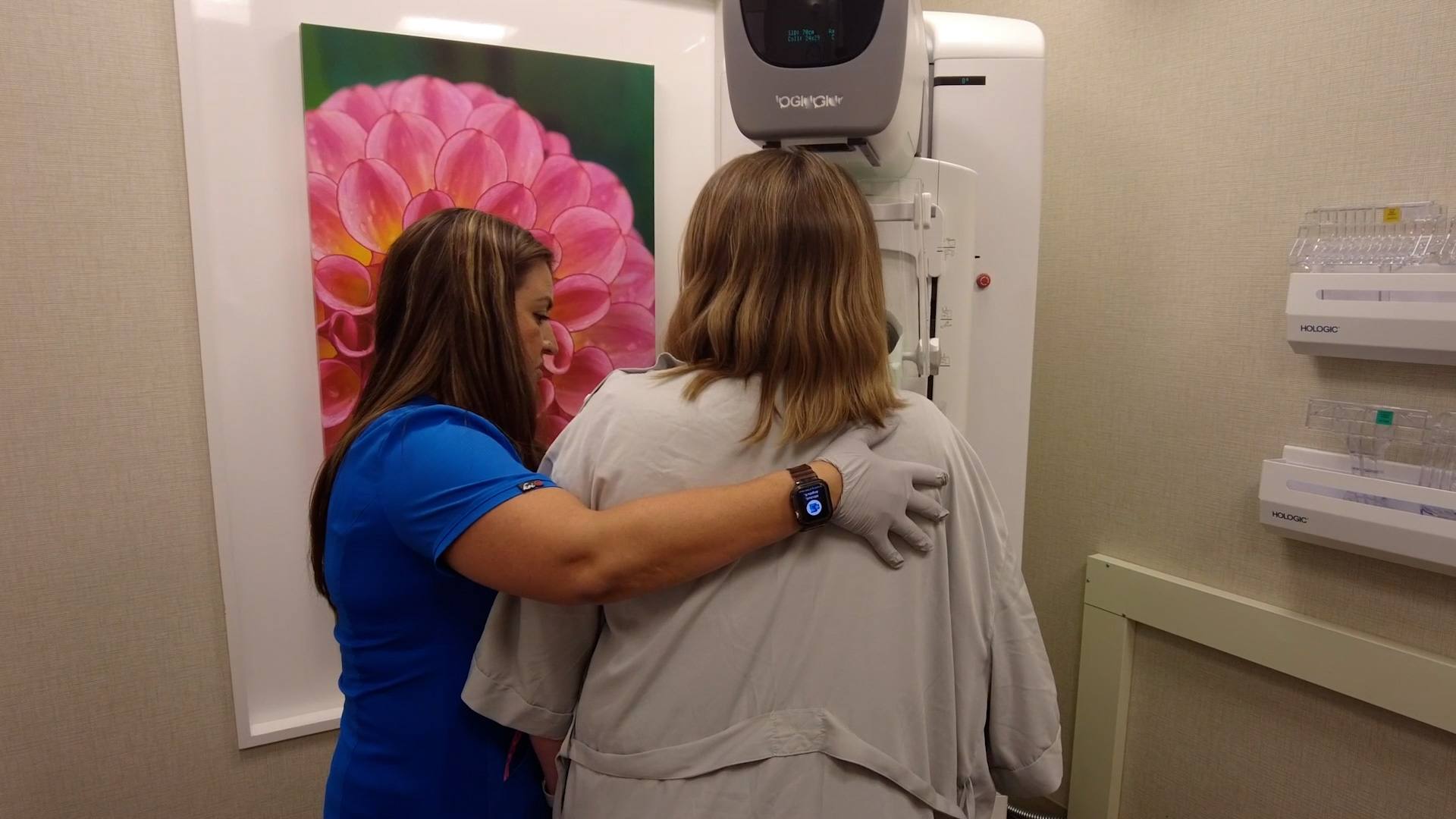
October is Breast Cancer Awareness Month. Breast cancer is the second-leading cause of cancer deaths in women across the U.S. And rates of the disease[...]
July 15, 2013
July 8, 2013
July 8, 2013
June 25, 2013
June 14, 2013
June 10, 2013
May 22, 2013
May 20, 2013
Explore more topics
 Sign up


Mayo Clinic Connect
An online patient support community